
The molar tooth—also known as Tooth #6 (First Molar)—plays a pivotal role in grinding food and maintaining the jaw and facial structure. When this key molar is lost, not only is chewing ability significantly reduced, but the risk of bone resorption, bite misalignment, and other oral diseases also increases. This article will help you clearly understand what the first molar is, the consequences of its loss, and reputable restoration methods, especially Dental Implant treatment at Nha khoa 3C.
What is the First Molar (Tooth #6)?
The First Molar (Tooth #6), in dental nomenclature, is located right behind the two premolars (Tooth #4 and #5) and before the second molar (Tooth #7). There are typically two first molars on each arch (upper and lower), totaling four in the mouth. The first molar has a strong root system and crown, with a broad, rugged chewing surface that aids in breaking down food right at the start of the digestive process.
Beyond chewing function, the first molar also plays a vital role in defining the bite (occlusion), stabilizing the jawbone structure, and contributing to the maintenance of facial contours. Therefore, losing the first molar creates a large gap, initiating a chain of negative reactions affecting oral and overall health.
How Does First Molar Loss Affect Health?
When you lose a first molar, it’s not simply the absence of a tooth—it’s the beginning of a chain of consequences that profoundly impact chewing function, bone structure, and the entire digestive system. Here are 5 main aspects illustrating the significant harm caused by “first molar loss.”
1. Severely Reduced Chewing Force
The first molar plays the dominant role in grinding food, accounting for up to 70% of the entire arch’s chewing power. When it is lost, most of the chewing pressure shifts to the remaining teeth—mostly the incisors and premolars, which do not have a sufficiently wide chewing surface. As a result, you are forced to chew longer and use uneven chewing forces, leading to improperly ground food, and easily causing jaw muscle fatigue, TMJ (Temporomandibular Joint) pain, and discomfort after every meal.
2. Impact on the Digestive System
Improperly chewed food forces the stomach and intestines to “overwork” to continue breaking it down with acid and digestive enzymes. This often leads to bloating, indigestion, and can even irritate the stomach lining in the long run. If this situation is repeated, the digestive system weakens, nutrient absorption is poor, leading to body fatigue, nutrient deficiency, and potential intestinal diseases.
3. Bone Resorption and Premature Aging
The alveolar bone requires chewing force to maintain its density and volume. When the first molar is lost, the surrounding jawbone area no longer receives mechanical stress from chewing, and the process of bone resorption begins. Over time, this bone shrinks, the face appears less firm, cheeks become sunken, wrinkles appear prematurely, and the jaw muscles weaken. At this stage, tooth restoration becomes more complex and costly due to the lack of supportive bone structure.
4. Bite Misalignment (Malocclusion)
The gap left by the missing molar allows adjacent teeth to drift, tilt, or super-erupt to fill the space. The opposing tooth also tends to over-erupt (extrude) seeking a counterpart. The result is bite misalignment, causing uneven tooth wear, TMJ pain, and an imbalance of chewing forces on both sides—further exacerbating the malocclusion.
5. Increased Risk of Other Oral Diseases
The space created by the missing molar easily becomes a “trap” for food and plaque, creating a favorable environment for bacteria to flourish. If not thoroughly cleaned, this gum area is prone to inflammation, bleeding, the formation of periodontal pockets, and decay in adjacent teeth. Periodontal inflammation not only destroys more alveolar bone but is also linked to systemic diseases like cardiovascular issues and diabetes.
Restoration Methods for a Missing First Molar
Dental Bridge
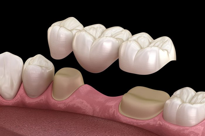
A dental bridge is a traditional restoration option where the dentist grinds down the two adjacent teeth to create two supporting abutments for one or two porcelain crowns spanning the missing space. Thanks to the quick procedure, after just a few appointments and about one to two weeks, the patient regains a continuous block of teeth, meeting basic chewing needs and providing immediate aesthetic improvement.
-
Pros: A dental bridge restores the tooth’s form with fairly natural color, reasonable cost, and does not require complex intervention on the jawbone.
-
Cons: Grinding down healthy teeth to create abutments significantly reduces natural tooth tissue, paving the way for sensitivity and dentin decay over time. Since the bridge lacks an artificial root, the underlying alveolar bone continues to resorb, leading to the risk of gum recession or exposure of the crown margin after a few years of use.
Doctors usually recommend dental bridges for those missing one or two consecutive molars, who have two strong natural adjacent teeth, stable jawbone structure, and need a quick solution with a moderate budget.
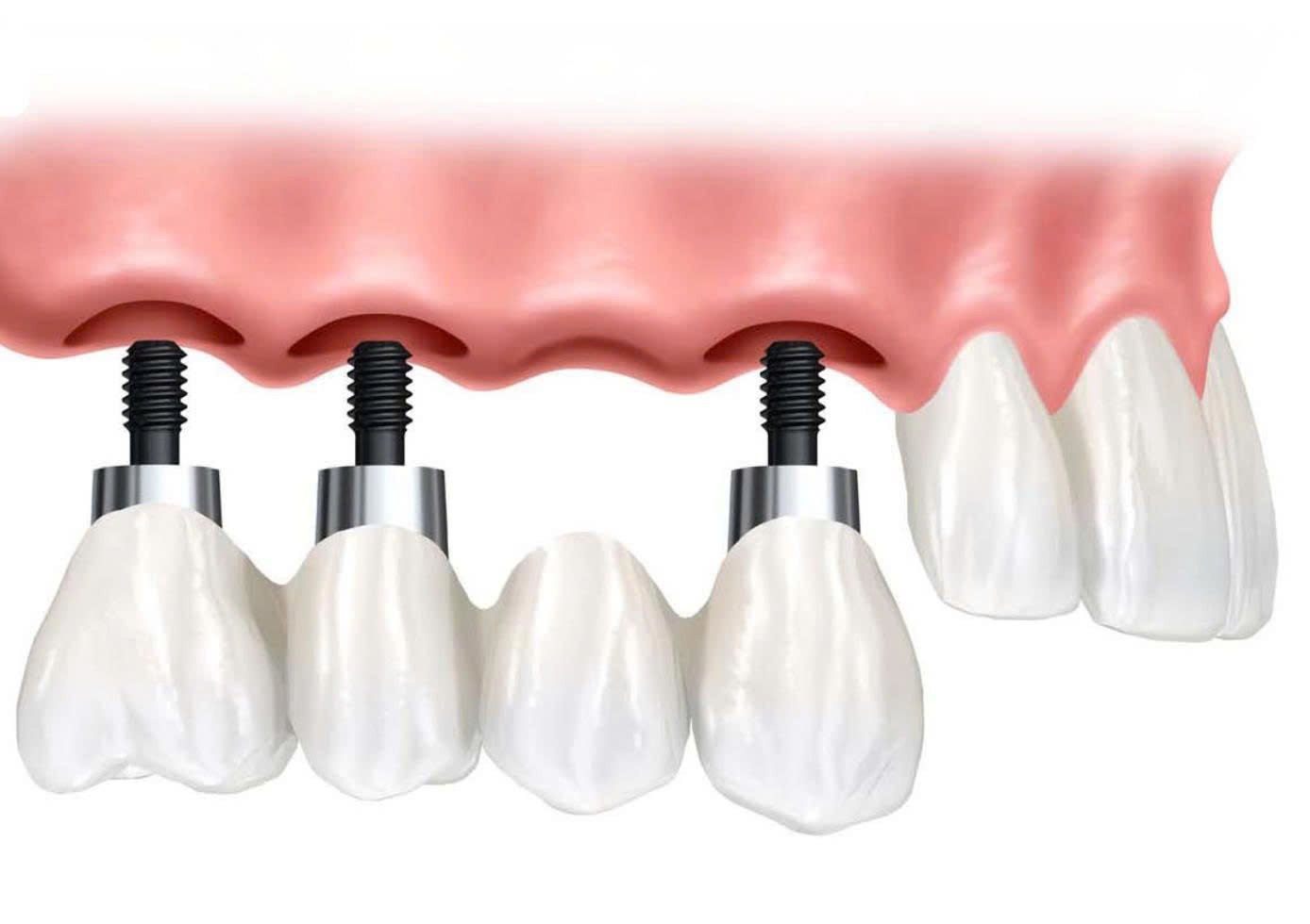
Dental Implant Placement
Dental Implant placement is the advanced technique that recreates both the tooth crown and the root by surgically placing a titanium post into the jawbone, followed by attaching an abutment and a porcelain crown. This approach not only fully restores chewing force—achieving over 90% of the natural grinding ability—but also ensures the alveolar bone is constantly under physiological stress, preventing bone resorption and slowing down facial aging. Aesthetically, the porcelain crown on an Implant is designed to fit snugly with the gum tissue, providing enduring, natural aesthetics over the years.
-
Cons: Implant placement requires a longer time for the post to integrate with the bone (ranging from three to six months), has a higher cost, and requires surgery with meticulous preparation steps, from 3D CT scans to assess bone density to supplemental bone regeneration when needed.
Doctors typically recommend Implants for patients with single or multiple tooth loss, stable overall health, sufficient jawbone height and width (or the potential for bone grafting), and those desiring a long-lasting solution that does not affect adjacent teeth.
Why Implant Placement Should Be Done Immediately After First Molar Loss
When the first molar is lost, the localized jawbone area immediately enters the bone resorption phase, while the gum tissue and gum line gradually deform. Immediate Implant placement—meaning placing the titanium post into the extraction socket during the same minor surgery—offers several important benefits. It not only helps the patient regain their tooth sooner but also optimizes function, aesthetics, and long-term economy.
1. Prevents Bone Resorption and Density Loss
Immediately after the first molar is lost, the bone destruction process begins intensely: within the first 3–6 months, alveolar bone volume can decrease by 40–60%, with the width particularly affected. By placing the Implant post right away, chewing forces and biological interaction are transmitted through the post to the bone, stimulating the bone remodeling mechanism and maintaining stable bone density. Thus, immediate Implant placement not only prevents bone volume loss but also preserves the height and width of the socket for subsequent restoration steps.
2. Preserves Soft Tissue and Maintains a Natural Gum Line
The gum tissue around the molar neck is delicate and prone to shrinking and recession if the extraction site is left empty for too long. Placing the Implant immediately helps the gum tissue adhere tightly around the post neck and supports the formation of the papilla—the small gum mounds between the teeth—which helps maintain a soft, natural gum line. In the long term, the patient avoids sunken cheeks, exposed Implant necks, or the “black margin” around the porcelain crown, preserving a natural appearance.
3. Shortens Treatment Time and Minimizes Bone Grafting
If the socket is left empty for a long time, the patient is often required to undergo bone grafting or guided bone regeneration (GBR) before Implant placement, which means extending the treatment time by an additional 3–6 months and incurring extra costs. Placing the Implant immediately after extraction utilizes the bone in the socket itself for post placement, limiting the need for supplemental bone grafting. Consequently, the total time to complete the restoration can be reduced by up to half, and the patient endures fewer surgeries and saves on treatment costs.
4. Improves Osseointegration and Implant Success Rate
Placing the Implant post into a fresh extraction socket often provides high initial stability (primary stability), as the post anchors tightly into the wall of the newly extracted socket, where the bone density is better than in bone that has already undergone resorption. This creates ideal conditions for effective osseointegration (bone integration), reducing the risk of failure due to a loose post or peri-implantitis. Furthermore, by minimizing scar tissue and infection, patients experience fewer complications, reduced pain, and a faster recovery time after surgery.
Dental Implant Placement at 3C Dental Clinic Helps Prevent Risks Associated with First Molar Loss
Highly Skilled and Experienced Doctor Team
At 3C Dental Clinic, every Implant placement case is performed directly by a team of doctors with over 10 years of specialized experience in Implantology and fixed restoration. With solid knowledge of jawbone anatomy, doctors accurately assess bone density and quality and select the appropriate surgical technique to minimize complications. Their rich clinical experience helps doctors flexibly handle all situations—from supplemental bone grafting to managing infections—while ensuring the Implant post integrates stably with a success rate over 99%.

Precision Guided Implant Technology
Utilizing 3D software and printed surgical guides, 3C Dental Clinic achieves the highest accuracy for post placement position. Before surgery, patients undergo a multi-dimensional CT Cone-Beam scan, and the data is transferred to the CAD/CAM system for virtual planning. The pre-printed surgical guide (from a 3D printer) helps the doctor insert the post at the correct angle and depth, avoiding encroachment on nerves and sinuses, reducing surgery time, and minimizing post-operative pain.
Immediate Loading Implant Technique for Chewing in 48 Hours
3C Dental Clinic is proud to implement the Immediate Loading Implant technique, allowing for the placement of a temporary tooth within 48 hours of Implant surgery. This method not only helps the patient eat sooner but also stimulates the osseointegration process, ensuring the most effective bone regeneration mobilization. Thanks to the temporary tooth being designed precisely for shape and bite, the functional recovery period is virtually uninterrupted, allowing patients to return to normal activities without long waiting times.
Modern Facilities, Advanced Equipment
Internationally standardized sterile operating rooms, 3D CT scanners, digital impression devices, and integrated CAD/CAM laboratory facilities are all invested in synchronously at 3C Dental Clinic. The closed sterile environment, along with the HEPA air filtration system, helps prevent infection throughout the surgical procedure. Furthermore, the in-house lab line with automated porcelain milling machines allows for the fabrication of sub-millimeter accurate porcelain crowns, ensuring stable bite and long-lasting natural aesthetics.
Genuine, High-Quality Implant Materials
We exclusively use genuine Implant posts imported from reputable dental corporations such as Straumann, Nobel Biocare, and Osstem. Medical-grade titanium (Grade 4) combined with SLA or SLActive treated surfaces enhances the osseointegration process and minimizes the risk of peri-implantitis. The porcelain crowns on the abutments are also selected from high-grade porcelain brands, being lead-free and metal-free, ensuring long-term mechanical durability and aesthetic brilliance.